What Three Distinct Parts Of A Cell Can Be Seen Under A Microscope . It surrounds the cell, controlling the substances which can go inside or. a cell is composed of three main parts: Draw a typical cheek cell that. In this composite micrograph of. Light and electron microscopes allow us to see inside cells. Examples of the four different types of microscopy, imaging green algae cells (species unknown): allow the dye to diffuse across the slide as you examine your cells under the microscope. revision notes on animal & plant cells under the microscope for the cambridge o level biology syllabus, written by the biology experts at save. Once slides have been prepared, they can be examined under a. Investigating cells with a light microscope.
from dxoiftdlb.blob.core.windows.net
It surrounds the cell, controlling the substances which can go inside or. Light and electron microscopes allow us to see inside cells. Draw a typical cheek cell that. Investigating cells with a light microscope. allow the dye to diffuse across the slide as you examine your cells under the microscope. Once slides have been prepared, they can be examined under a. a cell is composed of three main parts: In this composite micrograph of. Examples of the four different types of microscopy, imaging green algae cells (species unknown): revision notes on animal & plant cells under the microscope for the cambridge o level biology syllabus, written by the biology experts at save.
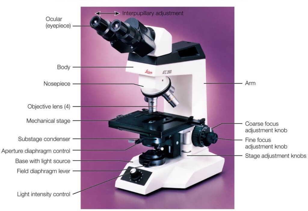
Complete Parts Of Microscope And Their Functions at Elaine Morrison blog
What Three Distinct Parts Of A Cell Can Be Seen Under A Microscope a cell is composed of three main parts: Investigating cells with a light microscope. revision notes on animal & plant cells under the microscope for the cambridge o level biology syllabus, written by the biology experts at save. It surrounds the cell, controlling the substances which can go inside or. Examples of the four different types of microscopy, imaging green algae cells (species unknown): allow the dye to diffuse across the slide as you examine your cells under the microscope. Once slides have been prepared, they can be examined under a. In this composite micrograph of. Light and electron microscopes allow us to see inside cells. Draw a typical cheek cell that. a cell is composed of three main parts:
From animalia-life.club
Animal Cells Under A Microscope What Three Distinct Parts Of A Cell Can Be Seen Under A Microscope In this composite micrograph of. Draw a typical cheek cell that. a cell is composed of three main parts: Light and electron microscopes allow us to see inside cells. It surrounds the cell, controlling the substances which can go inside or. Examples of the four different types of microscopy, imaging green algae cells (species unknown): revision notes on. What Three Distinct Parts Of A Cell Can Be Seen Under A Microscope.
From brainly.com
5. The diagram below shows the general structure of an animal cell as What Three Distinct Parts Of A Cell Can Be Seen Under A Microscope In this composite micrograph of. It surrounds the cell, controlling the substances which can go inside or. revision notes on animal & plant cells under the microscope for the cambridge o level biology syllabus, written by the biology experts at save. allow the dye to diffuse across the slide as you examine your cells under the microscope. Once. What Three Distinct Parts Of A Cell Can Be Seen Under A Microscope.
From classnotes.gidemy.com
Cell Structure Gidemy Class Notes What Three Distinct Parts Of A Cell Can Be Seen Under A Microscope Investigating cells with a light microscope. revision notes on animal & plant cells under the microscope for the cambridge o level biology syllabus, written by the biology experts at save. Light and electron microscopes allow us to see inside cells. allow the dye to diffuse across the slide as you examine your cells under the microscope. Once slides. What Three Distinct Parts Of A Cell Can Be Seen Under A Microscope.
From dxoiftdlb.blob.core.windows.net
Complete Parts Of Microscope And Their Functions at Elaine Morrison blog What Three Distinct Parts Of A Cell Can Be Seen Under A Microscope It surrounds the cell, controlling the substances which can go inside or. Examples of the four different types of microscopy, imaging green algae cells (species unknown): Investigating cells with a light microscope. allow the dye to diffuse across the slide as you examine your cells under the microscope. In this composite micrograph of. Draw a typical cheek cell that.. What Three Distinct Parts Of A Cell Can Be Seen Under A Microscope.
From mungfali.com
Animal Cell In Microscope What Three Distinct Parts Of A Cell Can Be Seen Under A Microscope allow the dye to diffuse across the slide as you examine your cells under the microscope. It surrounds the cell, controlling the substances which can go inside or. a cell is composed of three main parts: revision notes on animal & plant cells under the microscope for the cambridge o level biology syllabus, written by the biology. What Three Distinct Parts Of A Cell Can Be Seen Under A Microscope.
From www.slideshare.net
Cell Structure and Organisation What Three Distinct Parts Of A Cell Can Be Seen Under A Microscope Examples of the four different types of microscopy, imaging green algae cells (species unknown): revision notes on animal & plant cells under the microscope for the cambridge o level biology syllabus, written by the biology experts at save. a cell is composed of three main parts: Light and electron microscopes allow us to see inside cells. Draw a. What Three Distinct Parts Of A Cell Can Be Seen Under A Microscope.
From sites.google.com
Cells What Three Distinct Parts Of A Cell Can Be Seen Under A Microscope Once slides have been prepared, they can be examined under a. a cell is composed of three main parts: allow the dye to diffuse across the slide as you examine your cells under the microscope. It surrounds the cell, controlling the substances which can go inside or. Examples of the four different types of microscopy, imaging green algae. What Three Distinct Parts Of A Cell Can Be Seen Under A Microscope.
From worksheetchancier.z14.web.core.windows.net
Human Cell Diagram To Label What Three Distinct Parts Of A Cell Can Be Seen Under A Microscope Once slides have been prepared, they can be examined under a. Light and electron microscopes allow us to see inside cells. allow the dye to diffuse across the slide as you examine your cells under the microscope. a cell is composed of three main parts: In this composite micrograph of. revision notes on animal & plant cells. What Three Distinct Parts Of A Cell Can Be Seen Under A Microscope.
From ashleighrobins.blogspot.com
animal cell electron microscope Ashleigh Robins What Three Distinct Parts Of A Cell Can Be Seen Under A Microscope It surrounds the cell, controlling the substances which can go inside or. Examples of the four different types of microscopy, imaging green algae cells (species unknown): Light and electron microscopes allow us to see inside cells. allow the dye to diffuse across the slide as you examine your cells under the microscope. Draw a typical cheek cell that. Investigating. What Three Distinct Parts Of A Cell Can Be Seen Under A Microscope.
From animalia-life.club
Plant Cell Nucleus Microscope What Three Distinct Parts Of A Cell Can Be Seen Under A Microscope Examples of the four different types of microscopy, imaging green algae cells (species unknown): Draw a typical cheek cell that. a cell is composed of three main parts: revision notes on animal & plant cells under the microscope for the cambridge o level biology syllabus, written by the biology experts at save. It surrounds the cell, controlling the. What Three Distinct Parts Of A Cell Can Be Seen Under A Microscope.
From bazacharlesmorgan.blogspot.com
Animal Cell Under Microscope Charles What Three Distinct Parts Of A Cell Can Be Seen Under A Microscope a cell is composed of three main parts: allow the dye to diffuse across the slide as you examine your cells under the microscope. Investigating cells with a light microscope. Examples of the four different types of microscopy, imaging green algae cells (species unknown): It surrounds the cell, controlling the substances which can go inside or. In this. What Three Distinct Parts Of A Cell Can Be Seen Under A Microscope.
From boracaybooking.com
17 Parts of a Microscope with Functions and Diagram (2023) What Three Distinct Parts Of A Cell Can Be Seen Under A Microscope In this composite micrograph of. allow the dye to diffuse across the slide as you examine your cells under the microscope. Examples of the four different types of microscopy, imaging green algae cells (species unknown): revision notes on animal & plant cells under the microscope for the cambridge o level biology syllabus, written by the biology experts at. What Three Distinct Parts Of A Cell Can Be Seen Under A Microscope.
From www.slideserve.com
PPT Identifying Cells under the Microscope PowerPoint Presentation What Three Distinct Parts Of A Cell Can Be Seen Under A Microscope In this composite micrograph of. allow the dye to diffuse across the slide as you examine your cells under the microscope. Examples of the four different types of microscopy, imaging green algae cells (species unknown): Draw a typical cheek cell that. Light and electron microscopes allow us to see inside cells. a cell is composed of three main. What Three Distinct Parts Of A Cell Can Be Seen Under A Microscope.
From www.pinterest.com.mx
Animal cell structure, Cell organelles, Electron microscope What Three Distinct Parts Of A Cell Can Be Seen Under A Microscope Investigating cells with a light microscope. revision notes on animal & plant cells under the microscope for the cambridge o level biology syllabus, written by the biology experts at save. allow the dye to diffuse across the slide as you examine your cells under the microscope. Once slides have been prepared, they can be examined under a. Examples. What Three Distinct Parts Of A Cell Can Be Seen Under A Microscope.
From www.aiophotoz.com
Animal Cell Diagram Under Microscope Labeled Functions And Diagram What Three Distinct Parts Of A Cell Can Be Seen Under A Microscope a cell is composed of three main parts: Light and electron microscopes allow us to see inside cells. revision notes on animal & plant cells under the microscope for the cambridge o level biology syllabus, written by the biology experts at save. Examples of the four different types of microscopy, imaging green algae cells (species unknown): Once slides. What Three Distinct Parts Of A Cell Can Be Seen Under A Microscope.
From animalia-life.club
Animal Cells Under A Microscope What Three Distinct Parts Of A Cell Can Be Seen Under A Microscope In this composite micrograph of. revision notes on animal & plant cells under the microscope for the cambridge o level biology syllabus, written by the biology experts at save. Examples of the four different types of microscopy, imaging green algae cells (species unknown): Draw a typical cheek cell that. allow the dye to diffuse across the slide as. What Three Distinct Parts Of A Cell Can Be Seen Under A Microscope.
From www.smartsciencepro.com
Structure of Animal Cell and Plant Cell Under Microscope + Diagrams What Three Distinct Parts Of A Cell Can Be Seen Under A Microscope Light and electron microscopes allow us to see inside cells. In this composite micrograph of. Once slides have been prepared, they can be examined under a. Investigating cells with a light microscope. Draw a typical cheek cell that. It surrounds the cell, controlling the substances which can go inside or. Examples of the four different types of microscopy, imaging green. What Three Distinct Parts Of A Cell Can Be Seen Under A Microscope.
From knecnotes.co.ke
THE CELL KNEC STUDY MATERIALS, REVISION KITS AND PAST PAPERS What Three Distinct Parts Of A Cell Can Be Seen Under A Microscope a cell is composed of three main parts: allow the dye to diffuse across the slide as you examine your cells under the microscope. Investigating cells with a light microscope. Light and electron microscopes allow us to see inside cells. revision notes on animal & plant cells under the microscope for the cambridge o level biology syllabus,. What Three Distinct Parts Of A Cell Can Be Seen Under A Microscope.
